
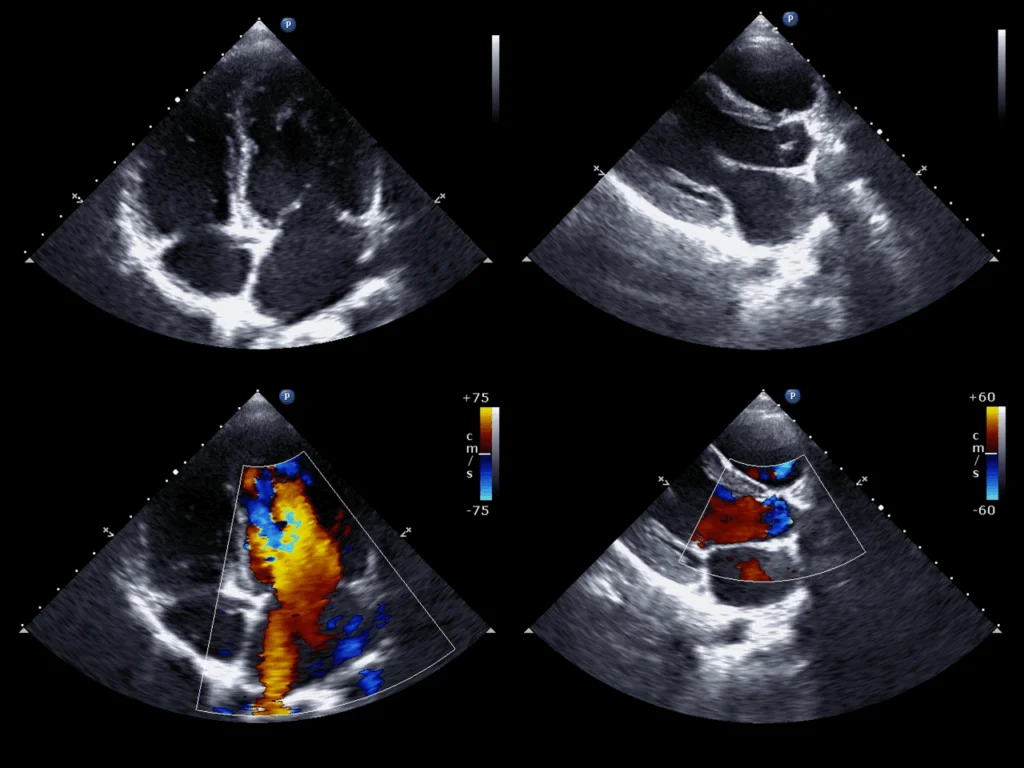
This test is valuable because it offers a complete evaluation of both heart structure and function. While 2D Echo shows chambers and pumping action, Colour Doppler reveals blood flow patterns, and Strain Imaging detects subtle dysfunction. Together, they enable early diagnosis of heart disease, guide treatment, and monitor therapy.
 Key Benefits and Applications
Provides detailed visualization of heart chambers, valves, and wall motion.
Detects valve leakage, narrowing, and abnormal blood flow patterns.
Identifies early ventricular dysfunction with strain imaging.
Useful in diagnosing and grading heart failure.
Assists in the management of congenital and acquired heart disease.
Tracks therapy response in chronic cardiac conditions.
More sensitive than conventional echocardiography alone for subtle abnormalities.
Non-invasive, safe, and repeatable test suitable for both routine and advanced cardiac care.